Brain Roads — Voxel Model, Datavisualisation Programme #5, 2020-2022
After having questioned the use of colour in tractographic representations of the brain, we question the formal register of the 3D models used and their adequacy to reality.
We find that the use of organically inspired 3D envelopes struggles to capture the different states of matter that surgeons encounter during operations. The tumour is not a clearly delimited foreign body; it is an alteration of the brain matter itself. This alteration is not uniform but progressive, making the tumour a diffuse entity that is difficult to circumscribe. Current images therefore pose a problem of accuracy of representation and can lead to misinterpretation. How then can we represent the uncertainty, the information actually available, without extrapolating our knowledge by using inadequate forms?
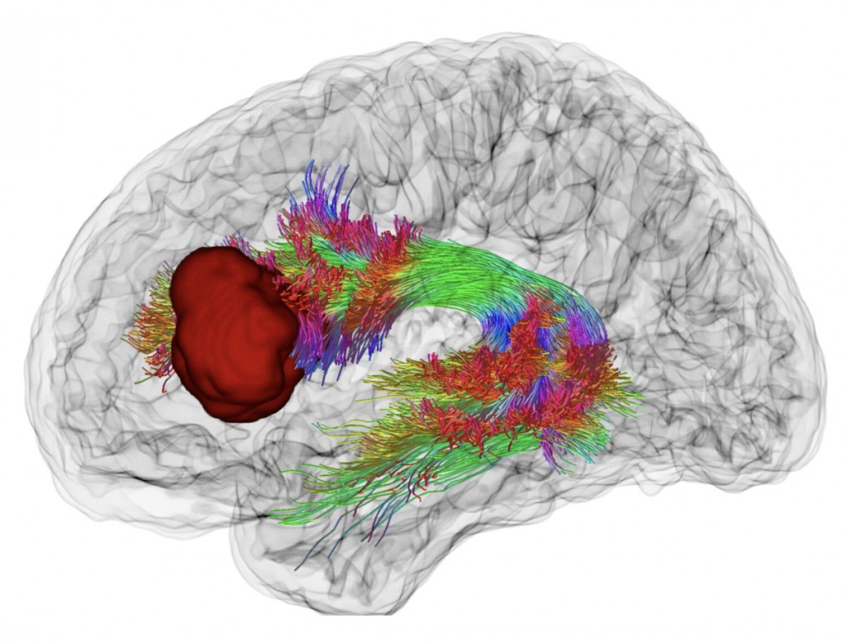
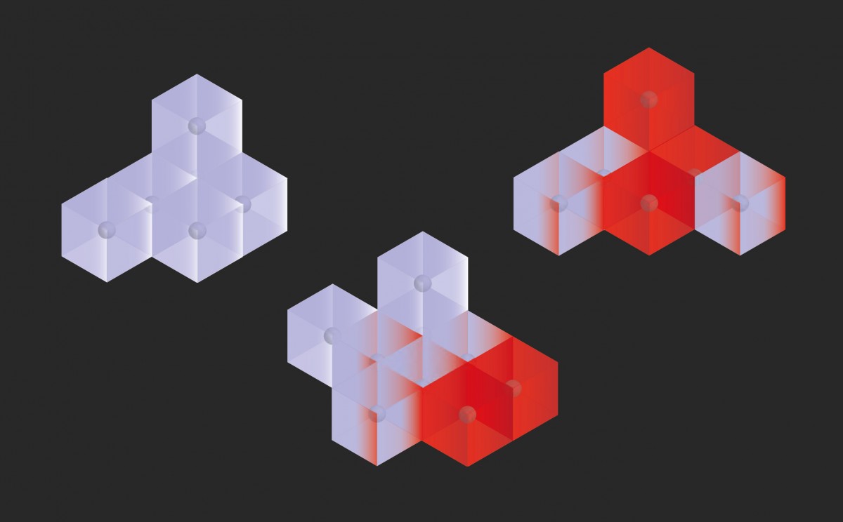
Current 3D bio-inspired shapes used to represent the brain and the tumor
Hypothesis
Our experimental attempt is to remove the organically inspired 3D shapes and return to a representation system based on the matrix of voxels - the volumetric pixels of the MRI scan - which constitute the real information available, and then to develop a meta-commentary capability on each voxel position. We hypothesise that this shift in the mode of representation allows us to move from realistic to real. By leaving the realism of 3D forms, we enter an aesthetically abstract model, a kind of mental space that expresses with the same intensity the recorded information and the unknown that surrounds it, thus offering a representation that is closer to the real level of information available on the observed brain.
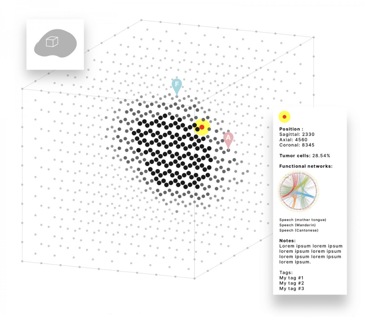
Outline of a voxel model applied to the representation of a brain. Olaf Avenati.
Seeing Through Digitised Matter
MRI scanned material produces a dataset of voxels: volumetric pixels, arranged in a matrix, which record a state of the material with coordinates. The voxel is therefore the smallest grain of information recorded. It is nevertheless relatively coarse (1mm on a side), in relation to the matter analysed (the brain). The voxel is usually represented as an opaque cube, which varies in colour. This is why MRIs are generally observed on screen in the form of successive slices to allow the complete information of the recording to be taken into account. The challenge for us is to analyse and work on the shape of the voxel to enable the volumetric aspect of the information to be perceived without necessarily using slices that would act as filters for the information.
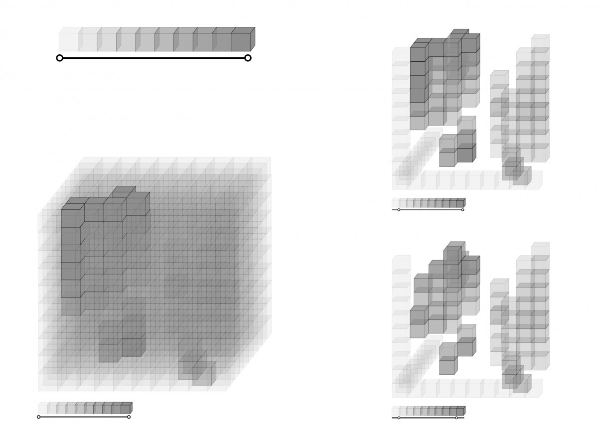
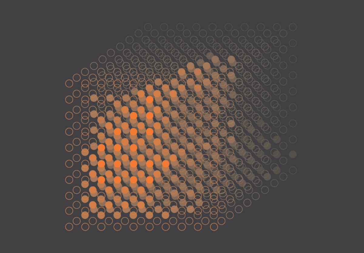
Filtering of voxels by applying value ranges. Graphical search. Antoine Sigur.
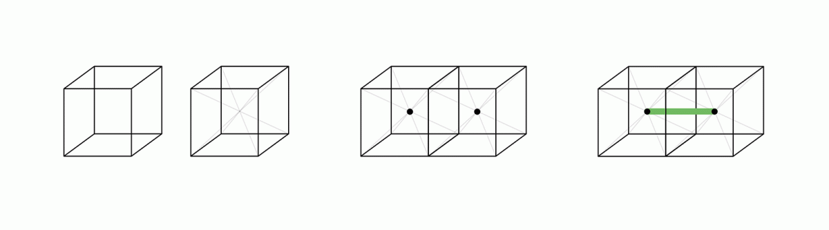
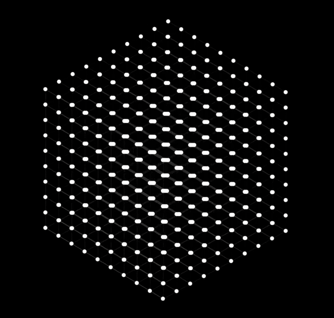
From Voxel-cube to Voxel-point: changing the representation convention to make the digitised material transparent without losing information along the way. Olaf Avenati.
From Voxel-cube to Voxel-point. Graphical research. Laurie Paolin.
Representing a voxel-point matrix. Graphical research. Amélie Blachère.
Setting things in motion
Evolution of a matrix of voxel-points. Animated experiment. Grégoire Gamichon & Charles Dessaint.
Modifier vos réglages de cookies
Dynamic filtering of a matrix of voxels. Animated experimentation with Cinema-4D software. Maia Xavier.
[ ongoing edition. Come back later to accès additional content ]
Partnership of four institutions
ESAD de Reims
Télécom SudParis - Institut polytechnique de Paris
Cluster of Excellence Matters of Activity, Humboldt-Universität zu Berlin - Image Space Material. Project: Adaptive Digital Twin
Charité-Universitätsmedizin Berlin (Hôpital Universitaire) - Image Guidance Lab. Department of Neurosurgery with Pediatric Neurosurgery
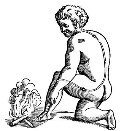
René Descartes, « Traité de l’homme », 1664. The long fiber from the foot to the head cavity is activated by the perception of heat and releases a fluid that contracts the muscles / La longue fibre qui va du pied à la cavité de la tête est actionnée par la perception de la chaleur et libère un fluide qui contracte les muscles. source
Principal investigators
Olaf Avenati - ESAD de Reims
Thomas Picht - Charité & Cluster MOA, HU Berlin
Patricia Ribault - Cluster MOA, HU Berlin
Michel Simatic - Télécom SudParis / IMT, IP Paris
Participants
FRANCE
ESAD de reims
Guillaume Andres, Amélie Bachère, Fiona Bosquier, Charles Dessaint, Grégoire Gamichon, Hoyam Mraizika, Laurie Paolin, Antoine Sigur, Maïa Xavier, Master1 students of graphic and digital design dept.
Télécom SudParis - Institut polytechnique de Paris
Julien Dock and Erwan Castioni, Master1 students of TSP – Institut Polytechnique de Paris
ESAD de Reims:
Olaf Avenati, Graphic & Digital Designer, Lecturer
Télécom SudParis - Institut polytechnique de Paris:
Michel Simatic, Engineer, Senior lecturer
GERMANY
Charité Universitäts-medizin Berlin:
Thomas Picht, PD Dr. Med. Neurosurgeon
Lucius Fekonja, Project Leader “Cutting”
Cluster Matters of Activity. image, space, material:
Patricia Ribault, Junior Professor
Maxime Le Calvé, Post-doctoral researcher
» Other projects of the Datavisualization Program at ESAD de Reims [in french]
